IMPLEMENTATION OF THE THREE FIELD ELECTRON WRAP-AROUND TECHNIQUE FOR EXTENSIVE RECURRENT CHEST WALL CARCINOMA: DOSIMETRIC AND CLINICAL CONSIDERATION
Mollye Norris, AS, RTRT, CMD
St. Luke’s Hospital
Kansas City, Missouri
INTRODUCTION
Radiation Therapy (RT)
Extensive recurrent chest wall carcinoma is a geometric challenge for the Radiation Oncology team. The direct skin involvement often extends from the sternum around to the posterior thorax. McNeely et al.1 and Leavitt et al.² have investigated the use of electron arc therapy for this condition. While electron arc therapy is desirable, unfortunately, most sites do not have this capability. Pezner et al.³ devised an alternative treatment using a combination of stationary photon and electron fields, referred to as the reverse hockey stick technique. This approach is simple, highly reproducible and delivers effective radiation therapy. The only limiting factor is the inability to use multiple electron energies to compensate for the varying chest wall thickness. The solution at our institution is to use multiple electron energies in a three field electron wrap-around technique. This technique successfully minimizes damage to healthy lung tissue which often times lies just a few millimeters under the diseased skin. When indicated, we use an anterior photon field to treat the clavicular, axillary and proximal arm region. Reproducible positioning to facilitate this wrap-around technique requires total support of the ipsilateral arm during the photon field treatment and partial arm support during the treatment of the three electron fields. Use of the standard L shaped arm board is recommended for the anterior photon field, but it is too bulky for use with the electron fields. To treat the electron field in the axillary fold region the electron cone must be in direct contact with the patient’s arm, thus the arm board must be removed.
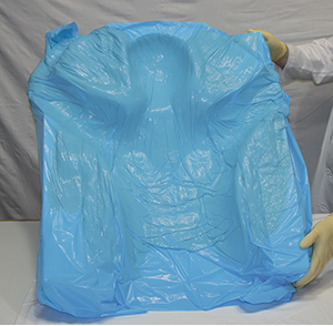
The success and precision of this treatment relies heavily on the use of a Smithers Medical Products, Inc. ALPHA CRADLE® brand Patient Repositioning System. The individualized, shaped upper body form provides continuous posterior proximal arm and elbow support. This immobilization device also ensures maximum patient comfort and precise daily reproducibility for a more efficient treatment. The cost for a customized ALPHA CRADLE® form is consistently less than 2% of the institutional charge for an average (22 intermediate treatments) radiation therapy program.
METHODS AND MATERIALS
Simulation
Positioning and form fabrication. The patient is first placed supine on the simulation table to determine the extent of arm mobility and optimal treatment position. The treatment requires that the patient’s ipsilateral arm be positioned as close as possible to a 90° angle from the lateral chest wall. The patient is then repositioned on the ALPHA CRADLE® MOLD MAKER II holding activated foaming agents. This system uses a slotted and beveled board with assorted dividers for creating forms for any anatomical site.4 At the time the foaming agents begin to expand and solidify it is important to elevate the elbow and the head slightly above the simulation couch. This enhances patient comfort and creates a stronger support form. Deep indentations are formed when the patient relaxes her head and arm into the partially solidified form. Since fabrication of a form takes approximately 15 minutes, it can easily be used with CT imaging immediately following simulation. Any excess piece of the form extending into the treatment region can easily be trimmed.
Portal verification. The photon-electron matchline must be slightly inferior to the axillary fold for consistency in matching. Using the physician’s premarked borders, the dosimetrist determines the length of the electron chest wall fields and marks the transverse central-axis of the anterior photon field is then positioned along the matchline with the central-axis being exactly halfway between the medial and lateral premarked borders. The inferior half of this field will be shielded with a split-beam block eliminating all divergence inferior to the matchplane. A verification radiograph of the anterior photon field is used by the radiation oncologist to indicate the appropriate customized blocking. In addition, a cross-table lateral radiograph is taken. This radiograph is used as a sagittal contour for external beam treatment planning to determine photon-electron gaps and appropriate junction moves via computer isodose distributions. For this radiograph, wires are placed on the patient’s anterior surface at the location of the central-axis of the photon field (not midline) extending inferiorly past the most inferior edge of the electron fields. At the completion of simulation, marks corresponding with alignment lasers can be added to the form for further accuracy.4
CT treatment planning
A transverse three slice CT scan is obtained with the patient in treatment position on the form. The CT flat couch insert is utilized for all CT radiotherapy treatment planning simulating the linear accelerator couch precisely. The patient is scanned at the level of the electron field central-axis, five centimeters inferior and five centimeters superior for three-dimensional visualization.
DOSIMETRIC COMPUTER PLANNING
Computer Algorithms
A dosimetrist rather than geometric method is used for determining the separation of and appropriate junction moves for adjacent fields. The placement of fields on the contour is optimized so that the composite isodose distribution is uniform at the desired depth and the ‘hot’ and ‘cold’ spots are acceptable. The accuracy of this procedure depends on the accuracy of the individual field isodose curves, especially in the penumbra region.5 A Computerized Medical Systems (CMS) radiotherapy treatment planning computer was used for all isodose calculations.6 It utilizes the pencil beam algorithm for calculation of electron beams.7,8 Comparison of measurement and calculated isodose curves revealed excellent agreement, with less than 3% or 3 mm of disagreement, except in the penumbra region at depth. The maximum variance was found in the 10% isodose line which exhibited an approximate 3 mm shift. Similar verifications have been reported by others.9 An analysis by Ibbott for high and low density inhomogeneities also verified the accuracy of CMS implementation of this pencil beam algorithm.9
The Clarkson integral algorithm is the calculational method of choice for extensively blocked photon beams. Hence, this algorithm was employed for the anterior photon field. Excellent agreement between measured and calculated isodose distributions have been verified, even in the presence of inhomogeneities.9
Three field electron wrap-around technique.
Chest wall. Utilization of CT interface has greatly enhanced radiotherapy treatment planning. Exact external contours, chest wall thicknesses and lung densities obtained through CT interface are critically important for precise dosimetry. Using such information, the dosimetrist performs computations on the transverse CT scan at the level of the central-axis of the electron fields using inhomogenity corrections. Because the skin lesions extend from the sternum around to the posterior thorax, three separate electron fields must be utilized. The goal of the dosimetrist is to keep the gantry angles as paralleled as possible to minimize hot and cold strips where the fields abut and diverge. Using a trial and error technique, the ideal gantry angles (approximately 10° increments), field sizes (dependent on chest wall thickness) are determined. Usually a 9 or 12 MeV electron beam or a combination of both energies proves successful. Electron field blocking is provided by three customized rectangularly shaped cerrobend (Lipowitz’s metal) shields, 9 mm thick. They are inserted into the electron cone applicator proximal to the patient’s skin for tertiary collimation.10 Additional lead malleable strips are carefully placed directly on the patient’s skin at all adjacent field borders to optimize electron beam definition.11 At the physician’s request, bolus material can be utilized to elevate the skin dose and minimize the dose gradient across the depth of the tumor volume.10 The bolus material also effectively eliminates the decreased dose at the skin surface within the gap regions.12
The D-max line has been normalized to 118% thereby placing the 80% depth isodose line at the pleural surface of the lung.³ These isodose curves reveal that a one-half centimeter gap is requited daily between the adjacent electron fields. This gap is determined by minimizing the dose gradient at the cross-field regions. To create a feathering effect of all overlap regions, a junction move is calculated by measuring the width of the hot strips (125%) between adjacent electron fields.
For this particular patient a one and one-half centimeter junction move is needed halfway through the treatment period. This junction move reduced the 125% hot to 120% and increased the cold strip from 80% to 90%, improving dose uniformity significantly. A composite computer plan is calculated, producing isodose distributions with appropriate bolus, gaps and junction moves. To minimize the frequency and severity of moist skin reaction, Pezner et al. recommend that the daily D-max dose to the electron fields not exceed 200 cGy.³
Anterior photon technique
Clavicular, axillary and upper arm. The anterior photon field uses elaborate custom blocking that depends heavily on daily reproducibility of the patient’s arm position. This position would be difficult to consistently reproduce without the custom made ALPHA CRADLE® form. An irregular field calculation is used to determine the blocked equivalent square and the axillary dose at midplane. A posterior axillary boost (PAB) could be added if indicated. Using the radiographic contour taken at the time of simulation, the sagittal isodose computer plan is generated. This plan demonstrates the need for a one-half centimeter gap between the photon-electron fields and a one centimeter junction move halfway through the treatment to create a feathering effect of all existing hot and cold strips.
CONCLUSION
This three field electron wrap-around technique is very useful in treating an extensive chest wall recurrence which extends from the sternum around to the posterior thorax when electron arc therapy is not available. An anterior photon field is implemented to treat skin lesions extending superiorly to the clavicle and along the proximal aspect of the arm. The success of this technique is due to the sophisticated treatment planning computer and algorithms, the skill of the treatment technologists, and the ALPHA CRADLE® custom form which provides precise reproducibility an maximum comfort for the patient.